

Removing Uraemic Toxins Through the Gut: The Enteric Dialysis Rationale in Azotaemia Management
A substantial share of uraemic toxins is produced not in the kidney but in the colon. The urea–ammonia cycle, protein-bound solutes and the gut–kidney axis: the basis and limits of the "enteric dialysis" rationale.
Summary / Direct answer: A substantial share of uraemic toxins is generated not in the kidney but in the large intestine, by bacterial metabolism. Urea diffuses into the gut lumen and is hydrolysed to ammonia by urease; indoxyl sulfate and p-cresyl sulfate arise from colonic fermentation of residual protein. "Enteric dialysis" is a metaphor for the nutritional approach targeting that intestinal source; it does not replace medical dialysis.
What is azotaemia, and what exactly do we mean by a uraemic toxin?
Azotaemia is the accumulation of nitrogenous waste products (urea nitrogen, creatinine) above the normal range. When it becomes clinically apparent — inappetence, nausea, vomiting, lethargy, uraemic halitosis, mucosal ulceration — we call it uraemia. In advanced renal disease, what challenges the clinician is rarely a single molecule; it is the sum of hundreds of solutes that accumulate as glomerular filtration rate declines.
These solutes fall broadly into three classes:
- Small water-soluble molecules: urea, creatinine, uric acid. Readily cleared by urine or dialysis.
- Middle molecules: peptides such as β2-microglobulin.
- Protein-bound solutes: indoxyl sulfate, p-cresyl sulfate, TMAO. Tightly bound to albumin, they are difficult to clear by filtration and even by haemodialysis.
The third group sits at the centre of the gut–kidney axis discussion, because their source is not the kidney at all. It is the colonic microbiota.
MECHANISM: how the gut–kidney axis works
1. Urea returns to the gut
Urea is not merely an end product excreted in urine; as it accumulates in blood it diffuses passively into the intestinal lumen along its concentration gradient. There, urease-producing bacteria hydrolyse it into ammonia and carbon dioxide. The resulting ammonia does two things: it raises luminal pH and it disrupts epithelial tight junction proteins (claudin-1, occludin, ZO-1). Experimental work by Vaziri and colleagues has linked this breakdown of the intestinal barrier in chronic kidney disease directly to the urea–ammonia cycle.
Once the barrier loosens, bacterial products and endotoxin leak into the systemic circulation, feeding low-grade chronic inflammation. One end of the inflammatory background so commonly seen in renal failure begins here.
2. Protein-bound toxins are manufactured in the colon
Protein that reaches the colon unabsorbed undergoes proteolytic rather than saccharolytic fermentation:
- Tryptophan → indole → (hepatic sulfation) → indoxyl sulfate
- Tyrosine / phenylalanine → p-cresol → p-cresyl sulfate
In experimental models both molecules have been associated with tubular cell stress, oxidative burden and endothelial dysfunction. In cats, studies have likewise reported a relationship between the faecal microbiome profile of individuals with chronic kidney disease and serum indoxyl sulfate and p-cresol sulfate concentrations.
The key point: these are molecules the kidney does not produce, yet is obliged to clear. When filtration capacity falls, the load rises — and one way to address the load is to look at where it is produced: the colon.
3. Uraemic dysbiosis closes the loop
In advanced renal disease the composition of the gut flora shifts: bacterial families producing urease, uricase, indole and p-cresol increase in relative terms, while saccharolytic species producing short-chain fatty acids (notably butyrate) recede. Because butyrate is the principal energy source of the colonocyte and a supporter of barrier integrity, this shift deepens the barrier problem. The result is a self-reinforcing loop: azotaemia → dysbiosis → barrier disruption → inflammation → greater toxin load.
4. Diverting nitrogen from urine to faeces
When fermentable fibre reaches the colon it provides substrate for the expansion of bacterial biomass. Proliferating bacteria incorporate luminal nitrogen into their own protein structures, and that nitrogen leaves the body with the faecal biomass. In human nephrology, fermentable fibre supplementation has been shown to increase faecal nitrogen excretion and to influence serum urea nitrogen. This is largely the physiological basis of the "enteric dialysis" metaphor: the gut lumen behaves like a second doorway through which nitrogen leaves the body.
Is "enteric dialysis" really dialysis?
No. The term is an analogy used to convey the concept, and it is used in this article in a metaphorical sense only. Haemodialysis and peritoneal dialysis are medical interventions that exchange solutes across a semipermeable membrane — the dialyser or the peritoneum — and require equipment, vascular or catheter access, anticoagulation and specialist supervision.
Nitrogen removal via the gut, by contrast, is not an intervention but the nutritional support of a physiological route that already exists. No complementary feed replaces dialysis, can claim to postpone it, or may be presented as an alternative to renal replacement therapy.
This must be stated plainly: diagnosis, staging and treatment planning are the exclusive remit of the veterinary surgeon. This article is a scientific review of mechanism, not clinical decision support.
Where does Azo-Fix sit in this picture?
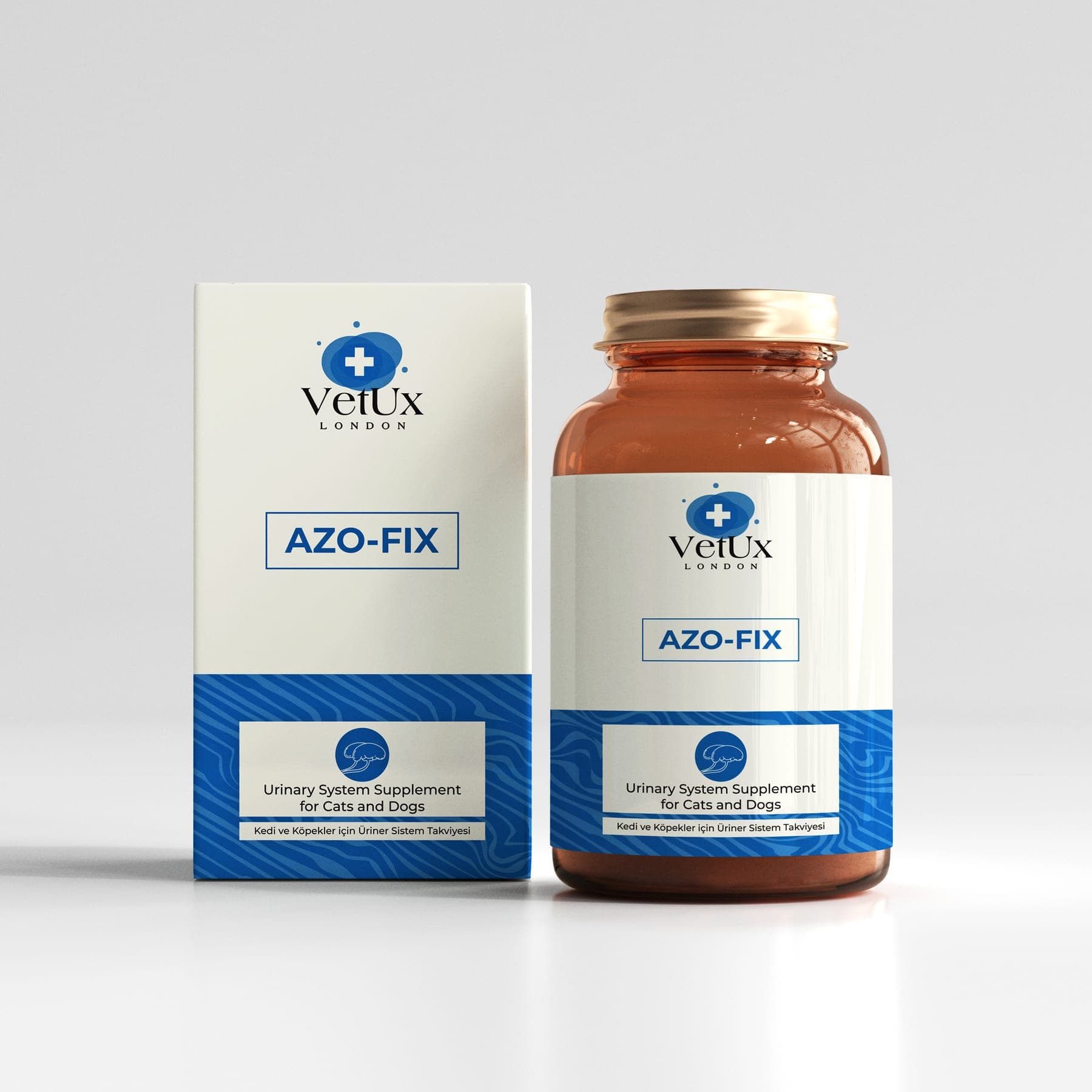
Azo-Fix is a powdered complementary feed for cats and dogs — not a medicine, not a treatment product. Its formulation is designed to give nutritional support to different links of the gut–kidney axis described above. The figures below are composition amounts per kilogram of product; they do not express any amount to be given to an animal.
Defined probiotic strains
The formula contains Enterococcus faecium NCIMB 10415 and Lactobacillus acidophilus CECT 4529 at 6.6 × 10¹² CFU per kilogram. Both are defined at strain level — the difference between saying "contains probiotics" and saying "contains which strain, at what viable count", and a decisive one in scientific assessment. Defined strains accompany the normal balance of the gut flora and the natural regulation of luminal urease activity.
Amino acids that enter nitrogen metabolism
- L-arginine and L-citrulline: two key intermediates of the urea cycle. Citrulline is converted via argininosuccinate to arginine; arginine then completes the cycle, releasing ornithine and urea. Arginine is essential in the cat; classic nutrition studies demonstrated that ammonia metabolism deteriorates rapidly in cats fed an arginine-free diet. These two amino acids therefore sit at the very centre of the natural process by which ammonia is converted into a less harmful metabolite.
- Taurine: a sulphur amino acid, essential in cats and conditionally essential in dogs, involved in bile salt conjugation and cellular osmoregulation.
- DL-methionine: as a sulphur amino acid it gives nutritional support to sulphur metabolism and to the normal physiological balance of urine.
- Vitamin B6 (pyridoxine HCl): cofactor of transamination reactions, accompanying the normal enzymatic steps in the handling of amino acid nitrogen.
Botanical components
- Cranberry (Vaccinium macrocarpon) — the highest-dosed botanical in the formula. Its proanthocyanidin content is among the most extensively studied botanical topics in the context of the natural balance of bacterial adhesion to urinary epithelium.
- Nettle leaf (Urtica dioica) and parsley (Petroselinum crispum) — plants traditionally associated with urinary system support, positioned here as components accompanying the normal course of fluid balance.
- Valerian (Valeriana officinalis) — contributes to the maintenance of calm. This is no minor detail: in cats in particular, stress directly affects urination behaviour and water intake.
- Pumpkin extract — as a prebiotic fibre source, it forms the nutritional leg of the nitrogen-diversion mechanism described above.
- Rosemary (Rosmarinus officinalis) and broccoli (Brassica oleracea) extracts — with their polyphenol and glucosinolate content, they accompany cellular antioxidant balance.
The product consists entirely of these components; the official reference for the formulation is the product's current record.
How should it be positioned in clinical practice?
Nutritional support does not replace the treatment plan built on IRIS staging; it accompanies it. The best-evidenced pillars of chronic kidney disease management in cats and dogs remain: an appropriate phosphorus-restricted renal diet, hydration management, control of hypertension and proteinuria, and regular monitoring.
A complementary feed should be considered within that framework, in addition to the plan set by the clinician. How and how often it is used is defined not by a product label but by the assessment of the veterinary surgeon examining the patient — which is why no amount is recommended in this article. It is used as advised by your veterinary surgeon.
What is the current state of the evidence?
Honesty is the only route to trust. The mechanism of the gut–kidney axis is robustly characterised in human nephrology, and a relationship between microbiota and protein-bound uraemic solutes has been demonstrated in cats as well. By contrast, data on the clinical effect of synbiotic support on azotaemia parameters in veterinary medicine are limited and conflicting. One feline study reported that an enteric-coated synbiotic product produced no change in azotaemia parameters when sprinkled onto food — with the discussion noting that loss of capsule integrity may have affected the outcome.
What this means in practice: the mechanism is sound, the clinical evidence is still maturing. The correct stance in the field is to position a complementary feed not as an item that "resolves azotaemia", but as a component giving nutritional support to the plan the clinician has established.
References
- 1Vaziri ND, Wong J, Pahl M, et al. Chronic kidney disease alters intestinal microbial flora. Kidney Int. 2013;83(2):308–315.
- 2Vaziri ND, Yuan J, Norris K. Role of urea in intestinal barrier dysfunction and disruption of epithelial tight junction in chronic kidney disease. Am J Nephrol. 2013;37(1):1–6.
- 3Evenepoel P, Meijers BKI, Bammens BRM, Verbeke K. Uremic toxins originating from colonic microbial metabolism. Kidney Int Suppl. 2009;(114):S12–S19.
- 4Meijers BKI, Evenepoel P. The gut–kidney axis: indoxyl sulfate, p-cresyl sulfate and CKD progression. Nephrol Dial Transplant. 2011;26(3):759–761.
- 5Niwa T. Indoxyl sulfate is a nephro-vascular toxin. J Ren Nutr. 2010;20(5 Suppl):S2–S6.
- 6Summers SC, Quimby JM, Isaiah A, et al. The fecal microbiome and serum concentrations of indoxyl sulfate and p-cresol sulfate in cats with chronic kidney disease. J Vet Intern Med. 2019;33(2):662–669.
- 7Rishniw M, Wynn SG. Azodyl, a synbiotic, fails to alter azotemia in cats with chronic kidney disease when sprinkled onto food. J Feline Med Surg. 2011;13(6):405–409.
- 8Ranganathan N, Ranganathan P, Friedman EA, et al. Pilot study of probiotic dietary supplementation for promoting healthy kidney function in patients with chronic kidney disease. Adv Ther. 2010;27(9):634–647.
- 9Bliss DZ, Stein TP, Schleifer CR, Settle RG. Supplementation with gum arabic fiber increases fecal nitrogen excretion and lowers serum urea nitrogen concentration in chronic renal failure patients consuming a low-protein diet. Am J Clin Nutr. 1996;63(3):392–398.
- 10Morris JG, Rogers QR. Arginine: an essential amino acid for the cat. J Nutr. 1978;108(12):1944–1953.
- 11Howell AB. Bioactive compounds in cranberries and their role in prevention of urinary tract infections. Mol Nutr Food Res. 2007;51(6):732–737.
- 12Sparkes AH, Caney S, Chalhoub S, et al. ISFM Consensus Guidelines on the Diagnosis and Management of Feline Chronic Kidney Disease. J Feline Med Surg. 2016;18(3):219–239.
- 13International Renal Interest Society (IRIS). Treatment Recommendations for CKD in Dogs and Cats. IRIS Staging Guidelines.
This content is provided for scientific information only and does not replace examination by a veterinary surgeon. Diagnosis and treatment are carried out by a veterinary surgeon alone. Azo-Fix is a complementary feed.
Frequently asked questions
Does Azo-Fix treat kidney failure?
No. Azo-Fix is a complementary feed; it is not a medicine and makes no treatment claim. In renal disease, diagnosis, staging and treatment decisions belong solely to the veterinary surgeon who examines the patient. A complementary feed is nutritional support accompanying the care that clinician has determined.
Does "enteric dialysis" mean my patient is being dialysed?
No. The expression is a metaphorical concept describing nitrogen removal via the gut. It has nothing to do with haemodialysis or peritoneal dialysis, does not replace them and does not remove the need for them.
Can it be used alongside a renal diet?
Complementary feeds are considered alongside, never instead of, the clinical nutrition plan. Your veterinary surgeon makes the holistic assessment of your patient's diet, additional support and concurrent medication; the decision to combine them is theirs.
How much is given, and how?
No amount is given in this article. The manner, frequency and duration of use vary with the patient's stage, weight, appetite and concurrent treatment; it is used **as advised by your veterinary surgeon.**
Why does it matter that the probiotic strain is defined?
Probiotic effects are assessed at strain level, not species level. Strains identified by collection numbers such as NCIMB 10415 and CECT 4529 make it traceable which microorganism is supplied and at what viable count — the first criterion to look for in scientific assessment and in product comparison.
Can it be used in a healthy animal?
This article is not a usage recommendation. Whether any supplement is appropriate for a healthy or a diseased animal is decided by a veterinary surgeon following examination and laboratory assessment.
This article is for general information only; it does not replace veterinary examination, diagnosis or treatment. VetUx London products are complementary feed. If you have any concern about your companion, please consult your veterinary surgeon.
Share
The formulas matching the nutritional support described above.
Related articles
All articles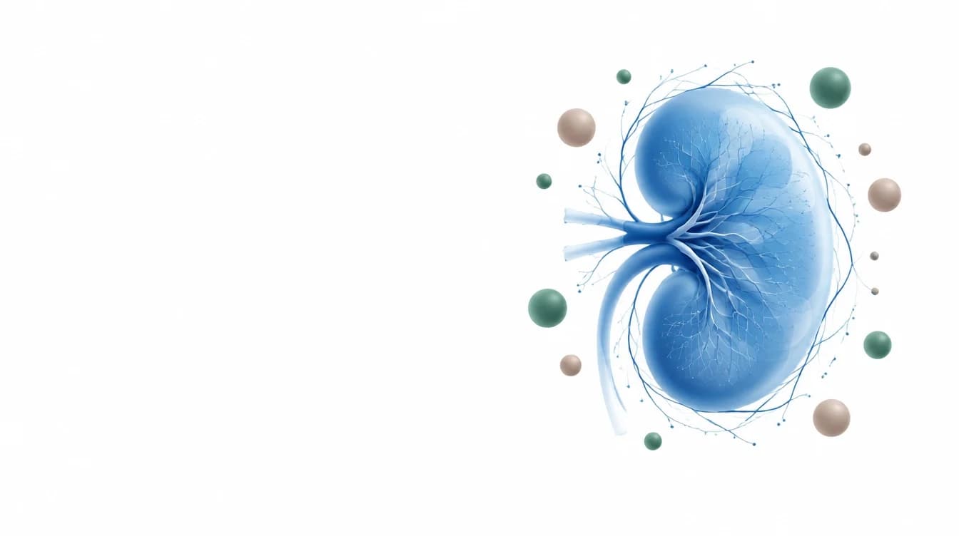 Clinical Notes
Why Are Phosphorus Retention and Microcirculation Vital in Chronic Kidney Disease?
In chronic kidney disease, blood phosphorus can look normal until the adaptive capacity of the remaining nephrons is exhausted. We explain the FGF-23–PTH axis and renal microcirculation at the level of general physiology.
 Clinical Notes
What Do Raised ALT and AST Mean in Cats and Dogs? The Hepatoprotective Role of Silymarin and Artichoke
Raised ALT and AST are not a diagnosis but a laboratory finding your vet reads. We look at why these enzymes leak into the blood, the described membrane mechanisms of silymarin, and the role of artichoke extract in bile flow — with the limits of the evidence stated plainly.
 Clinical Notes
Do Hairballs Irritate a Cat's Stomach? Gastritis Risks and Scientific Approach
Accumulation of trichobezoars in cats mechanically erodes the gastric mucosa, predisposing to chronic gastritis. Nutritional and clinical approaches for physiological transit.