
Suture Site Healing in Cats and Dogs: Scarring, Granulation Tissue and Topical Protection
A suture site heals through haemostasis, inflammation, proliferation and remodelling. We explain what is normal at home, how collagen organisation shapes a scar and where topical protection fits; diagnosis and treatment belong to your veterinary surgeon.
Short answer: In cats and dogs, a suture site heals in four phases: haemostasis, inflammation, proliferation (granulation and epithelialisation) and remodelling. The skin surface closes within the first days, while collagen fibres reorganise over months. Keeping the area clean, dry and protected from licking is essential; if you see redness, discharge or the wound opening, contact your veterinary surgeon.
Neutering, lump removal or orthopaedic surgery: whatever the procedure, the suture line is the first thing an owner checks once home. "Is this redness normal?", "Why hasn't the fur grown back?", "Will there be a scar?" These questions all relate to a highly ordered biological programme that the skin runs at a millimetre scale. In this article I walk through that programme phase by phase, explain what to watch for at home, and look at the place of topical protection in the light of the evidence.
How does a suture site heal? The mechanism of the four phases
Wound healing consists of four overlapping phases. They are less a calendar than a chain of cellular events, each one triggering the next (Hosgood, 2006; Gurtner et al., 2008).
1. Haemostasis (first minutes)
As soon as the incision is made, vessels constrict, platelets aggregate and a fibrin mesh forms. Beyond stopping bleeding, this clot acts as a temporary scaffold. Growth factors released by platelets (PDGF, TGF-β) recruit the cells of the next phases.
2. Inflammation (roughly the first 3–5 days)
Neutrophils arrive first and clear bacteria and debris. Macrophages follow. The macrophage is often called the "conductor" of healing, because it both cleans the wound and releases signals that stimulate fibroblasts and vascular cells. During this period, mild, symmetrical pinkness and slight swelling at the wound edges can be physiological. Redness that increases or spreads, warmth, or discharge is not physiological inflammation and should be assessed by your veterinary surgeon.
3. Proliferation: granulation tissue and epithelialisation (from about day 3)
- Granulation tissue: Fibroblasts multiply and initially produce mainly type III collagen. New capillaries (angiogenesis) give this tissue its red, velvety, "granular" appearance. Granulation tissue is a living scaffold that fills the wound space.
- Epithelialisation: Keratinocytes at the wound edges migrate across the surface to close it. A surgical incision whose edges have been brought together (primary closure) is usually bridged superficially by a thin layer of epithelium within the first 24–48 hours. Epithelialisation progresses faster on a moist, protected surface than under a dry scab (Winter, 1962).
- Contraction: Myofibroblasts pull the wound edges towards each other. This is most noticeable in open wounds that heal by second intention.
4. Remodelling (from weeks to a year or more)
In a suture line that looks "closed", the real work happens in this phase. Type III collagen is replaced by thicker, stronger type I collagen. Fibres are reinforced by cross-links, and matrix metalloproteinases break down and reorganise disordered fibres. In the first weeks the wound has only a small fraction of the strength of intact skin. Strength increases over months but never fully returns; at most it reaches about 70–80% of intact skin (Gurtner et al., 2008).
What is granulation tissue, and why is there so little in a sutured wound?
Granulation tissue is a natural and necessary stage of healing. How much of it you see depends on how the wound was closed:
| | Primary healing (sutured incision) | Second-intention healing (wound left open) | |---|---|---| | Edges | Brought together | Open, with a gap | | Granulation tissue | Minimal, usually not visible | Prominent, fills the defect | | Epithelialisation distance | Millimetres | Can be centimetres | | Scar | A fine line | Wider |
Cats are not small dogs. Experimental studies comparing healing in cats and dogs have shown that in cats granulation tissue forms later and in smaller amounts, and that epithelialisation and contraction are slower. The breaking strength of sutured wounds at one week was also markedly lower in cats than in dogs (Bohling et al., 2004). The same researchers reported that the subcutaneous tissues contribute less to the blood supply of the wound in cats (Bohling et al., 2006). In practice this means that a feline incision that looks fine on the surface may not yet be fully strong underneath. This is why your veterinary surgeon decides when sutures come out and when exercise restrictions are lifted.
How does collagen fibre organisation shape the appearance of a scar?
In intact skin, collagen fibres form a "basket-weave" network running in different directions. In scar tissue, the fibres are denser, more parallel and less elastic. Skin appendages such as hair follicles and sweat glands generally do not regenerate. A mature scar may therefore remain as a fine, pale, hairless line.
The main factors that impair collagen organisation and scar quality:
- Tension and movement: Incisions over joints or in areas under tension tend to leave wider scars.
- Licking, scratching and rubbing: Repeated mechanical trauma prolongs the inflammatory phase.
- Infection and seroma: These set healing back. Prospective surveillance in dogs has identified surgical site infections after discharge from the clinic (Turk et al., 2015), which is why observation at home matters so much.
- Systemic factors: Diabetes, hyperadrenocorticism, long-term glucocorticoid use, protein deficiency and obesity can slow healing.
When will the fur grow back? Regrowth in a clipped area depends on the hair cycle and can take months, especially in double-coated breeds (Diaz et al., 2006). Directly on the suture line there are no follicles left, so fur may never regrow there. This is not necessarily a sign of a problem. If the hairless area is spreading, or there is scaling or itching, consult your veterinary surgeon.
How should I monitor the suture site at home?
Looking at the suture line every day, at the same time and in good light, is the easiest way to spot changes early.
| Usually expected | Contact your veterinary surgeon if you see | |---|---| | Mild, symmetrical pinkness at the edges in the first days | Redness that increases or spreads, or warmth in the area | | Slight swelling that gradually decreases | Swelling that grows, whether soft or firm | | A small amount of clear seepage on the first day | Cloudy, yellow-green, foul-smelling or bloody discharge | | A dry line with edges touching | Opening of the wound edges, or broken or missing sutures | | Increasing comfort day by day | Pain, lethargy, loss of appetite, fever |
⚠️ If you notice redness, discharge or opening at the suture site, contact your veterinary surgeon before doing anything at home. Unless your vet recommends it, do not apply hydrogen peroxide, alcohol or home remedies to the area; these can damage healing tissue.
Your veterinary surgeon will also decide whether a protective collar or surgical body suit is needed to prevent licking.
What does topical protection do, and what does it not do?
Keeping the skin surface clean, protected from external factors and at a balanced moisture level accompanies the physiological course of epithelialisation (Winter, 1962). This surface environment is where topical care products have their role: barrier, moisture balance and surface hygiene.
Topical care does not:
- replace surgical technique, suture care or veterinary check-ups;
- resolve infection, a reopened incision or a deep tissue problem; their diagnosis and treatment belong to the veterinary surgeon;
- remove a permanent scar. A scar is the natural result of the way skin repairs itself.
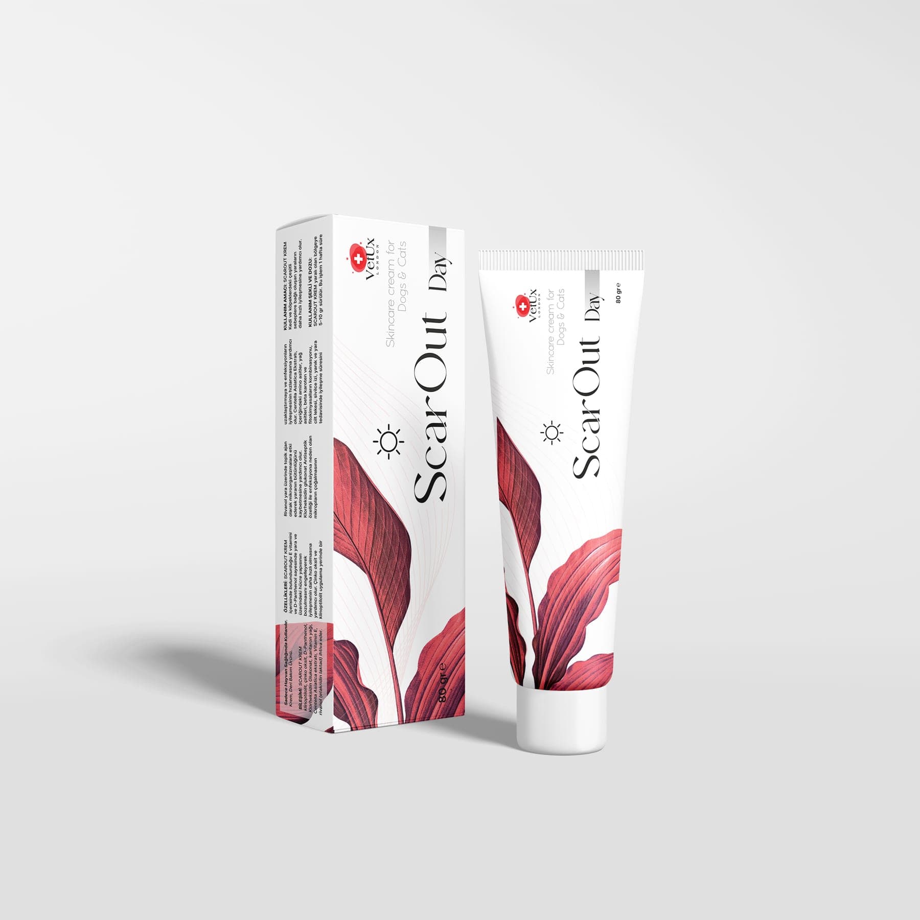
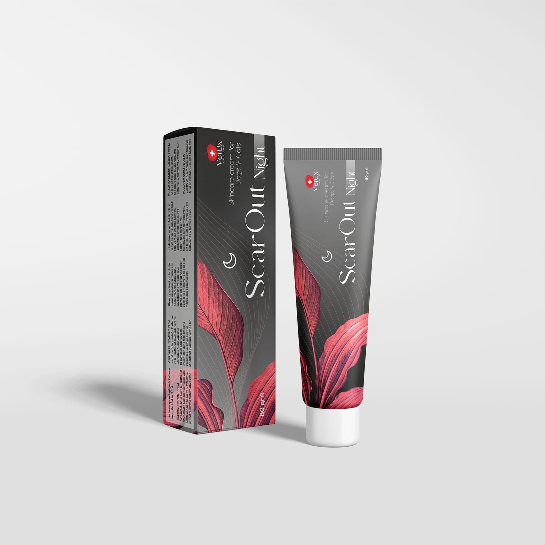
How do the ingredients of ScarOut Day and ScarOut Night accompany this process?
ScarOut Day and ScarOut Night are care creams for external use only, prepared for the care of the skin surface in cats and dogs. Both products have the same composition. They differ in texture: Day has a light texture suited to daytime use, while Night is richer, stays in place for longer and forms a covering layer through the night.
| Ingredient | General role in the literature | Care function in the product | |---|---|---| | Clinoptilolite (natural zeolite) | Porous, adsorbent mineral (Mastinu et al., 2019) | Helps keep the surface dry and protected | | Zinc oxide | Protective barrier; zinc is involved in the physiology of epithelial renewal (Lansdown et al., 2007) | Surface protection | | Centella asiatica extract | Its triterpenes have been reported in laboratory studies to accompany collagen synthesis (Bonté et al., 1994; Bylka et al., 2013) | Accompanies the natural process of collagen formation | | D-Panthenol (provitamin B5) | Epidermal barrier and moisture balance (Ebner et al., 2002) | Contributes to moisture balance | | Chlorhexidine gluconate | Broad-spectrum antiseptic (Lozier et al., 1992) | Contributes to surface hygiene | | Rivanol (ethacridine lactate) | Acridine-derived antiseptic | Contributes to surface hygiene | | St John's wort oil (Hypericum perforatum) | Traditional botanical oil studied in experimental wound models (Süntar et al., 2010) | Soothes skin that has become reactive | | Vitamin C (ascorbic acid) | Cofactor in collagen synthesis; antioxidant (Pullar et al., 2017) | Supports normal oxidative balance | | Vitamin E (tocopherol) | Lipophilic antioxidant; human data on its effect on scar appearance are conflicting (Baumann & Spencer, 1999) | Supports normal oxidative balance |
A note on the table: This information is drawn from the general scientific literature on the individual ingredients and is not an efficacy claim for the finished product. For transparency: ScarOut does not contain silicone gel, antibiotics, honey, hyaluronic acid or growth factors.
Usage warnings from the product record:
- For external use only.
- Apply to the cleaned skin surface, in consultation with your veterinary surgeon.
- Prevent the animal from licking the area.
- Store in a cool, dry place out of the reach of children.
- The record states that, where needed, the two products can be used together in a day-and-night routine.
Timing of application: The product record gives no specific information on use on a fresh suture line or an open wound. Your veterinary surgeon therefore decides when to start using the product (before or after suture removal) and on which area. Follow your vet's recommendation on how often to apply it.
Important note
This article is for information only and does not replace an examination by a veterinary surgeon. Diagnosis and treatment are the responsibility of the veterinary surgeon. ScarOut Day and ScarOut Night are care products for external use that accompany the care recommended by your veterinary surgeon. They are not intended to treat any disease.
References
- 1Hosgood G. Stages of wound healing and their clinical relevance. Vet Clin North Am Small Anim Pract. 2006;36(4):667–685. doi:10.1016/j.cvsm.2006.02.006
- 2Gurtner GC, Werner S, Barrandon Y, Longaker MT. Wound repair and regeneration. Nature. 2008;453(7193):314–321. doi:10.1038/nature07039
- 3Bohling MW, Henderson RA, Swaim SF, Kincaid SA, Wright JC. Cutaneous wound healing in the cat: a macroscopic description and comparison with cutaneous wound healing in the dog. Vet Surg. 2004;33(6):579–587. doi:10.1111/j.1532-950X.2004.04081.x
- 4Bohling MW, Henderson RA, Swaim SF, et al. Comparison of the role of the subcutaneous tissues in cutaneous wound healing in the dog and cat. Vet Surg. 2006;35(1):3–14. doi:10.1111/j.1532-950X.2005.00105.x
- 5Winter GD. Formation of the scab and the rate of epithelization of superficial wounds in the skin of the young domestic pig. Nature. 1962;193:293–294. doi:10.1038/193293a0
- 6Turk R, Singh A, Weese JS. Prospective surgical site infection surveillance in dogs. Vet Surg. 2015;44(1):2–8. doi:10.1111/j.1532-950X.2014.12267.x
- 7Diaz SF, Torres SMF, Nogueira SAF, Gilbert S, Jessen CR. The impact of body site, topical melatonin and brushing on hair regrowth after clipping normal Siberian Husky dogs. Vet Dermatol. 2006;17(1):45–50. doi:10.1111/j.1365-3164.2005.00493.x
- 8Lansdown ABG, Mirastschijski U, Stubbs N, Scanlon E, Ågren MS. Zinc in wound healing: theoretical, experimental, and clinical aspects. Wound Repair Regen. 2007;15(1):2–16. doi:10.1111/j.1524-475X.2006.00179.x
- 9Mastinu A, Kumar A, Maccarinelli G, et al. Zeolite clinoptilolite: therapeutic virtues of an ancient mineral. Molecules. 2019;24(8):1517. doi:10.3390/molecules24081517
- 10Bonté F, Dumas M, Chaudagne C, Meybeck A. Influence of asiatic acid, madecassic acid, and asiaticoside on human collagen I synthesis. Planta Med. 1994;60(2):133–135. doi:10.1055/s-2006-959434
- 11Bylka W, Znajdek-Awiżeń P, Studzińska-Sroka E, Brzezińska M. Centella asiatica in cosmetology. Postepy Dermatol Alergol. 2013;30(1):46–49. doi:10.5114/pdia.2013.33378
- 12Ebner F, Heller A, Rippke F, Tausch I. Topical use of dexpanthenol in skin disorders. Am J Clin Dermatol. 2002;3(6):427–433. doi:10.2165/00128071-200203060-00005
- 13Lozier S, Pope E, Berg J. Effects of four preparations of 0.05% chlorhexidine diacetate on wound healing in dogs. Vet Surg. 1992;21(2):107–112. doi:10.1111/j.1532-950X.1992.tb00025.x
- 14Süntar IP, Akkol EK, Yılmazer D, et al. Investigations on the in vivo wound healing potential of Hypericum perforatum L. J Ethnopharmacol. 2010;127(2):468–477. doi:10.1016/j.jep.2009.10.011
- 15Pullar JM, Carr AC, Vissers MCM. The roles of vitamin C in skin health. Nutrients. 2017;9(8):866. doi:10.3390/nu9080866
- 16Baumann LS, Spencer J. The effects of topical vitamin E on the cosmetic appearance of scars. Dermatol Surg. 1999;25(4):311–315. doi:10.1046/j.1524-4725.1999.08223.x
- 17Williams J, Moores A (eds). BSAVA Manual of Canine and Feline Wound Management and Reconstruction. 2nd ed. Gloucester: BSAVA; 2009.
Frequently asked questions
When are sutures removed?
Skin sutures are often removed at around 10–14 days. The timing depends on the location and tension of the incision, the species (healing can be slower in cats) and overall health, and your veterinary surgeon will set the date. Intradermal (absorbable) sutures may not need removing.
Is mild pinkness along the suture edges normal?
In the first few days, mild and symmetrical pinkness can be part of the inflammatory phase. Redness that increases or spreads, warmth, or discharge is not normal; contact your veterinary surgeon without delay.
Will the scar disappear completely?
No. A scar is the permanent result of skin being repaired with collagen. Over time it matures, becomes finer and fades, a process that can take up to a year. Topical care supports and protects the physiological renewal of the skin surface, but it does not remove scar tissue.
Can ScarOut be applied to fresh sutures?
The product record contains no specific instructions for fresh sutures or open wounds. Your veterinary surgeon decides when and where to apply it. The product is for external use only, on a cleaned skin surface, and licking should be prevented.
My cat or dog keeps licking the sutures. What should I do?
Licking causes mechanical trauma, carries oral bacteria to the area and can make the wound open. Ask your veterinary surgeon about a protective collar or surgical body suit. If the incision has opened, or discharge or redness has started, contact your vet.
Why is the fur slow to grow back over the surgical site?
Regrowth in a clipped area depends on the hair cycle and can take months. Directly on the suture line there are no follicles, so a fine hairless line may remain. If the hairless area is widening, a dermatological assessment may be needed.
This article is for general information only; it does not replace veterinary examination, diagnosis or treatment. VetUx London products are complementary feed. If you have any concern about your companion, please consult your veterinary surgeon.
Share
The formulas matching the nutritional support described above.
Related articles
All articles Clinical Notes
Gut Development in Pregnant Mothers and Nursing Young: The Move From Mother's Milk to Solid Food
Why do newborn kittens and puppies gain their immunity from colostrum, when does gut closure happen, and how should the move from mother's milk to solid food be planned? The physiology of nutrition through pregnancy and nursing; every decision belongs to your vet.
 Clinical Notes
Red Blood Cell Losses in Cats and Dogs: Bioavailability of Chelated Iron and Vitamin B12 Absorption
Why do red blood cell losses and anaemia of chronic disease cause lethargy in cats and dogs? Explore the hepcidin-ferroportin axis, the bioavailability of chelated iron and vitamin B12 (cobalamin) absorption, and the physiological principles of bone marrow erythropoiesis support.
 Clinical Notes
Hard Stools and Straining in Cats: The Peristaltic Power of Soluble and Insoluble Fibre
Why do cats develop hard stools and straining, and when does chronic constipation turn into a megacolon risk? This article covers the role of soluble and insoluble fibre in faecal bulk, water holding, peristalsis and the microbiota, plus the warning signs that need a veterinary surgeon.